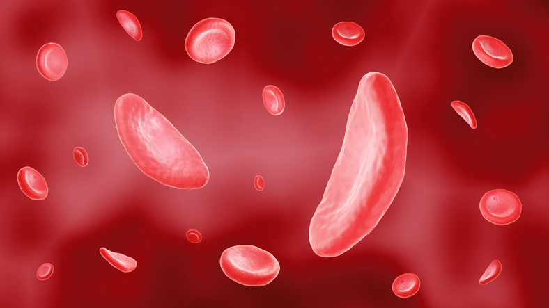
Phosphatidylserine (PS) and phosphatidylethanolamine (PE) are phospholipids that normally reside in the inner leaflet of the red blood cell membrane. Their presence on the outer membrane, which is cell-age dependent, contributes to hemolysis, clotting and immune activation. In patients with sickle cell disease (SCD), studies suggest the presence of PS and PE on the surface of RBCs may lead to increased vaso-occlusion events. To understand how surface PS and PE affect RBC transfusion outcomes in patients with SCD, researchers randomized 26 chronically transfused patients with SCD to receive either RBCs stored for <10 days or >30 days for three consecutive transfusion events and measured the amount of exposed PE and PS on the donor RBCs along with several markers of recipient transfusion efficiency (including hemoglobin [Hb]), the amount of sickled hemoglobin, hemolysis and others) 2-hours, 24-hours and 2-weeks after transfusion. While PS expression levels on donor RBCs were similar in the two study arms (4.6% vs 5.3% in the <10-day and >30-day arms, respectively), the level of PE surface expression was significantly higher in the >30-day arm (4.6% vs 8.5% for <10-day and >30-day arms, respectively). Irrespective of study arm, donor RBCs with high surface levels of PE and PS resulted in higher Hb increments compared to pre-transfusion levels at 24-hours and 2-weeks after transfusion (p=0.02 and p=0.04, respectively). Importantly, 86% (12/14) of severe adverse events requiring hospitalization occurred after transfusion of at least one RBC unit with high PE and PS surface expression; furthermore, all 27 non-severe pain-related adverse events occurred after transfusion of RBCs with high PE and PS surface expression. Further research is needed to better understand the relationship between aging cells and PS-PE surface expression.
Reference: